- Еуромедик, Булевар уметности 29, Београд

What is hamartoma?
Др Мерима Горан
Hamatromas are mostly benign changes that represent mature but architecturally incompletely organized tissues in a location that normally contains these tissues. Although many classify them as tumor like changes, because they actually represent malformations in development, they can still be true neoplastic lesions, considering that chromosomal aberrations occur through somatic mutations.
The term hamartoma was first used by Arrigoni et al. in 1971.
What is the frequency of occurrence of hamartomas?
Breast hamartomas are classified as benign tumors with an incidence of 0.7 – 4.8%. Their incidence is increasing, but it is primarily a reflection of the use of screening for breast cancer, when these lesions are diagnosed more often. Therefore, these changes are mostly an incidental finding. Although the incidence is apparently increasing, these tumors are still very rare and are described in the literature as single cases or case series.
Although a few cases have been recorded in men, these changes are generally more common in women, mostly middle-aged, while they are extremely rare in the pediatric (prepubescent) population.
How does it manifest itself?
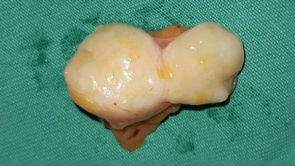
Clinically, they present as a slow-growing, solid tumor mass in the breast, oval to round in shape, mobile, clearly demarcated, with a smooth surface. They can be of different dimensions, from smaller non-palpable, accidentally discovered changes, up to large palpable masses in the breast even over 10 cm.
What are they made of?
Histologically, it is a benign lesion made of epithelial, fibrous and fatty tissue in different proportions, and since these tissues are integral elements of the breast, hamartomas are often called “a breast within a breast”. Depending on which component predominates, different terms are used to describe these changes: lipofibroadenomas, fibroadenolipomas, or adenolipomas.
Myoid hamartoma is a rare form that contains smooth muscle tissue in addition to fatty tissue. Also, microcalcifications and ossification can exist in these tumors.
Why do they arise?
The etiopathogenesis is not well known, but as mentioned, it is believed to be a dysgenesis rather than a true tumor. Female hormones are cited as possible etiopathogenetic factors, given that the expression of estrogen and progesterone receptors has been proven on certain tumors. The smooth muscle component in myoid hamartomas is likely to originate from the vessel wall, mammary, mammary myoepithelial cells, or undifferentiated stromal cells. Metaplasia of stromal cells to muscle cells is also a possible mechanism of origin, because CD4 +, SMA +, as well as the expression of desmin and vimentin have been demonstrated in myoid hamartomas immunohistochemically.
Hamartomas are a common finding in Cowden syndrome, which is a rare autosomal dominant inherited syndrome caused by a mutation of the PTEN tumor suppressor gene. It is characterized by the appearance of multiple hamartomas of the skin, mucous membranes, brain, breast, thyroid and gastrointestinal tract, abnormalities of the CNS, as well as an increased risk of developing breast and thyroid carcinoma compared to the general population.
Are they dangerous?
Cases of the development of invasive carcinoma in hamartoma have also been described. Malignant transformation is possible due to the presence of an epithelial component in the tumor, which leads to the development of cancer through cellular atypia. In terms of differential diagnosis, these tumors can be confused with fibroadenomas, but there is no fatty tissue in them.
How do they find out?
Ultrasound shows these changes as solid, clearly defined, avascular, inhomogeneous with hypo- and hyperechoic fields, depending on the amount of glandular, fibrous and fatty tissue. Mammographically, they appear as oval or round lesions, inhomogeneous with radiopaque and transparent fields depending on the structure, clearly limited by a thin capsule. If they have a large amount of fibrous tissue, then they have a more homogeneous structure and are difficult to distinguish from fibroadenomas, and if they have a large amount of fatty tissue, they are difficult to distinguish from lipomas. On MRI, these changes are shown as heterogeneous, and after the application of contrast, they show a mostly benign, type 1 curve.
How are they treated?
The therapy of choice is operative treatment, surgical excision of the entire tumor. Enucleation of these tumors is usually easy because they are mostly encapsulated or pseudocapsulated. Recurrences are rare after surgical excision.
Литература
1. Amir RA, Sheikh SS. Breast hamartoma: A report of 14 cases of an under-recognized and under-reported entity. Int J Surg Case Rep. 2016; 22:1-4.
2. Bhatia M, Ravikumar R, Maurya VK, Rai R. “Breast within a breast” sign: Mammary hamartoma. Med J Armed Forces India. 2015 Oct; 71(4): 377-9.
3. Presazzi A, Di Giulio G, Calliada F. Breast hamartoma: ultrasound, elastosonographic, and mammographic features. Mini pictorial essay. J Ultrasound. 2015 Jul 26; 18(4): 373-7.
4. Sevim Y, Kocaay AF, Eker T, Celasin H, Karabork A, Erden E, Genc V. Breast hamartoma: a clinicopathologic analysis of 27 cases and a literature review. Clinics (Sao Paulo). 2014 Aug; 69(8):515-23.
5. Gupta SS, Singh O, Hastir A, Arora G, Sabharwal G, Mishra H. Breast hamartoma with intrathoracic extension in a 13-year-old boy. J Cancer Res Ther. 2010 Jan-Mar; 6(1): 86-8.